한국의 동충하초 서론 동충하초란 쓰는 즐거움(2)
2021년 05월 13일. 오늘도 우리 남은 인생의 첫날을 맞이하는 목요일로 이제는 날씨도 좋아 들판이 농작물로 채워지는 풍성한 날을 맞이하면서 성재모동충하초를 벗 삼아 어디에 내어놓아도 손색이 없을 정도로 잘 재배하여 인연이 있는 고객님을 맞이하려고 한다. 언제나 고객님에게 고마운 마음을 드리며 이렇게 글을 올리는 것만으로도 기적이고 축복으로 알고 걸림이 없는 하루를 보내려고 한다. 오늘 또 하루를 선물로 받았네요.
오늘은 동충하초에 관한 구조에 대하여 글을 쓰는 시간을 가지었다. 이 세상에 존재하는 모든 생물은 참 복잡한 구조로 되어 있다. 동충하초는 작고 미생물에 속하므로 구조가 복잡하지 않으리라 생각하였는데 알면 알수록 복잡하다. 동충하초를 연구하면서 보이는 세계보다 보이지 않은 세계가 더 중요하다는 것을 알았다. 사람도 몸보다는 마음이 중요한 것처럼 말이다. 동충하초의 가장 중요한 포자는 육안으로는 볼 수 없고 포자가 곤충에 도달하면 발아하여 들어가서 균사체를 만들고 이 균사체는 곤충의 유충이 풀을 먹으면 그 풀을 이용하여 생장한다. 곤충의 몸을 다 채운 다음 마지막으로 동충하초가 몸을 뚫고 나온다. 우리 눈으로 볼 수 있는 것은 종류에 따라 다르지만 1달 정도이다. 지금 이 세상을 떠들썩하게 하는 코로나19도 보인다면 문제가 없을 텐데 보이지 않으니 속수무책으로 당하고 있다. 이런 면에서 사람들은 조금 겸손하여야 한다고 본다는 왜냐하면 바이러스 하나에 온 세상이 떠들썩하여도 이 바이러스를 제거하는 데는 오랜 세월이 지나가도 미생물을 연구한 나로서는 요원하다고 본다. 이제 미생물과 함께 순응하면서 살 수밖에 없다고 본다.
오늘은 동충하초의 구조 사진을 올려놓고 내가 맞이하는 모든 것에 순응하면서 오늘도 바르고 천천히 흔들림이 없이 그냥 가려고 한다. 우리 모두 사는 동안 최선을 다해 후회 없는 삶을 살았음 좋겠습니다. 그저 물처럼 흐르는 인생으로 늘 행복하고 웃음 가득 찬 나날이 되기를 기원합시다. 더욱 건강하시고, 범사가 잘 되는 복을 누리시기를 기원합니다. 고맙고 고맙습니다.
문의 : 010-4872-5936
주문 :
성재모동충하초 머쉬텍 제품 http://naver.me/5rLTrzqo
성재모동충하초 현미밥알칩 http://me2.do/F36egE3F
성재모동충하초 현미강정 http://naver.me/GUvhYm9X
성재모동충하초 주문서 서울개미 문소라 http://naver.me/FWJm0n0a
하심정 www.hsinsam.com
횡성새말양봉 www.smhoney.kr
횡성군 농업인백화점 https://cafe.naver.com/farmworkers/162
동충하초 http://sungjaemo.com http://blog.daum.net/cordyceps
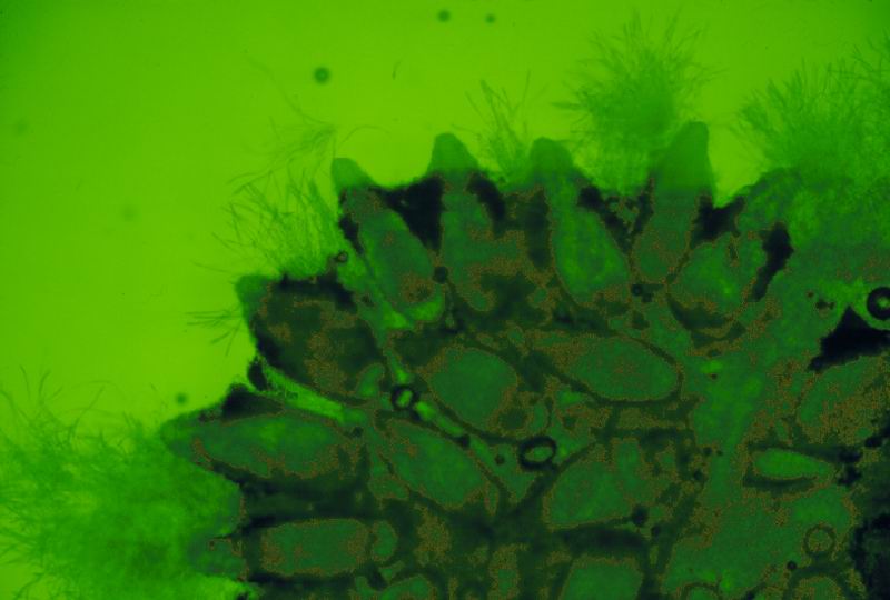
균학 용어에 있어서 동충하초 균에 속하는 균은 자낭균문과 접합균문에 속하며 특히 기주의 생활사 선택성이 있다. 예를 들면 그들은 주로 유충일 때 침입하나 간혹 번데기 혹은 성충에도 감염이 된다. 곤충기생균인 Cordyceps 균은 그들이 공격한 곤충의 몸 밖으로 한 개 혹은 여러 개의 자실체를 형성한다(그림 I-1 1). 동충하초는 동충하초균이 곤충을 침입하여 곤충을 죽게 한 다음, 기주(寄主)로부터 자좌(子座)를 형성하는 버섯을 말한다. 그러나 균이 곤충을 침입하여 곤충을 죽게 할지라도, 자좌를 형성하지 않고 기주의 표면에 무성생식(無性生殖) 기관인 분생포자(分生胞子)를 형성하는 종류도 있다. 곤충을 침입하는 대개의 곤충기생균은 자좌를 형성하지 않으며, 분생포자를 생산하는 것들이 대부분인데, 이들의 분류는 분생포자의 형태, 즉 분생포자가 분생자경 또는 작은 자루(小柄)에 접착한 형태에 따라 분류한다.
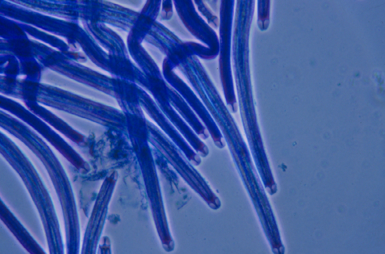
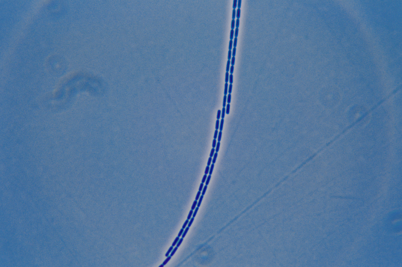
동충하초균은 자좌는 머리와 자루로 구성되어 있고(그림. I-1 2, 3, 4, 5, 6, 7) 땅속이나 썩은 나무 나뭇가지에 형성되는 곤충에서 나온 자좌는 자낭포자가 발산할 수 있도록 지상부로 나온다. 자좌는 머리 표면에는 알맹이 모양의 자낭각을 형성하고, 자낭각 내에는 주머니 모양의 자낭이 일렬로 배열되어 있다. 자낭 내에는 실 모양의 자낭포자를 형성한다. (그림. I-1 5). 자낭포자가 성숙하면 선단의 자낭구멍이 열려 공중에 포자를 비산(飛散)하여 곤충의 몸에 접착하게 된다. 동충하초는 자좌, 자낭각, 자낭, 자낭포자로 구성되었고 자낭에는 8개의 자낭포자가 들어있다. (그림. I-1 5). 곤충에서 형성된 자낭과 자낭포자가 성숙이 되면 자낭각의 정단에 있는 구멍을 통하여 자낭으로부터 자낭포자가 사출되어 곤충의 기주에 접착하여 발아한 후 효소를 내어 곤충의 조직을 뚫고 들어가 동충하초가 그 속에서 자라게 되어 다음 해 다시 동충하초를 형성하는 생활사를 시작한다. (그림. I-1 8). 동충하초균의 분류는 기주 이외에 자좌의 모양, 머리의 모양, 머리가 자루에 접착한 형태, 자낭각, 자낭과 자낭포자의 모양, 자낭포자가 2차 포자로 분열하는지 등이 주요한 분류 기준이 되고 있다.
(Figs. I-1 2, 3, 4, 5, 6, 7) 기주로부터 올라온 자루는 땅속에 묻혀 있거나, 썩은 나무, 나뭇잎의 표면에 있고 자낭포자가 형성되어 외부로 발산할 수 있도록 (Fig. I-1 5). (Fig. I-1 5). (Fig. I-1 8).
Fig. I-1. 동충하초의 구조 1, 유츙에 형성된 자좌 2, 반묻힌형자낭각 3, 자낭각 4, 자낭각의 내부. 5, 두꺼운 정단 뚜껑을 가진 길고 가는 자낭. 6, 자낭포자 7, 원형의 2차포자. 8, 자낭포자의 발아
In mycological terms, the fungi covered here belong to two main groups of fungi, Ascomycota and Zygomycota. Some of these fungi show considerable specificity for particular life stages of their hosts, e.g., they may infect only larvae, only pupae, or only adults of a single insect species. Insect-born fungi, in the case of Cordyceps species, form fruiting bodies (stromata; singular: stroma) outside of the bodies of the insects they attack (Fig. I-1 1). Many other forms of entomopathogenic fungi, most especially the conidial fungi, do not form stromata but do form conidia on hyphae on the surface of the insect host or on erect fascicles of hyphae called synnemata. Their conidial states associated with the entomopathogenic ascomycetes are classified according to the shapes of conidiogenous cells, the manner in which conidia are produced from those cells, and the shapes of those conidia.
The stromata of these fungi consist of a fertile part (or head) in which the perithecia, asci, and ascospores are produced (Figs. I-1, 2, 3, 4, 5, 6, 7) and a sterile stipe that supports the fertile part. The stipes, arising from affected hosts, buried in the ground, in rotting wood or under the surface of leaf litter, are long enough to assure that the ascospores are produced and discharged from an aerial position that greatly assists the ability of fungi to disperse effectively and to reach uninfected individuals of susceptible host species. Flask-like perithecia form on or partially to fully immersed in the surface of the fertile part. Inside these perithecia, asci (elongated cells with a characteristic cap-like apical thickening) are arranged in a row (Fig. I-1 5). Meiotic divisions (reduction divisions) take place only in asci, and after meiosis takes place, resultant nuclei may undergo one or more rounds of mitotic divisions before the cell walls that delimit the ascospores are laid down around one or more of these nuclei. In Cordyceps species and most other ascomycetes, eight ascospores are formed in each ascus (Fig. I-1 5). When ascospores and the asci in which they form are fully mature, the ascospores are forcibly dispersed as the asci literally squirt out the spores through the apical openings (ostioles) of the perithecia and then become airborne and may adhere to the body of another susceptible host insect to begin the cycle over again, following their germination (Fig. I-1 8). The classification of Cordyceps is the host, the shape of stipe, the shape of the head, the shape of attached head with stipe, perithecium, the shape of ascus and ascospore and seondary spore divided by ascospore.


'동충하초 책 > 한국동충하초(총론)' 카테고리의 다른 글
| 한국의 동충하초 서론에서 자좌에 형성되는 자낭 머리의 위치 모양 (0) | 2021.05.16 |
|---|---|
| 한국의 동충하초 서론에서 자좌의 모양을 살펴본다. (0) | 2021.05.15 |
| 한국의 동충하초 서론 동충하초란 쓰는 즐거움(1) (0) | 2021.05.12 |
| 한국의 동충하초 책 머리말을 쓰는 즐거움 (0) | 2021.05.10 |
| 한국의 동충하초 개정판을 내고 싶다. (0) | 2021.05.09 |